Facility
Kit's Endo is fully equipped with the latest advanced technology to help us in all phases of treatment, from diagnosis to root canal filling and sealing.
Below is a list and description of some of the most important pieces of equipment we use:
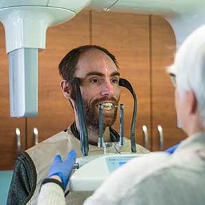
Computer Digital Radiography (CDR)
Dental x-rays can now be taken using sensors that transmit the image directly into a computer monitor. This larger image helps the patient understand the doctor's explanations more easily and enables the doctor to "zoom in" on a specific area of the tooth. An important advantage to this new technology is that it reduces radiation exposure. Digital x-rays are also faster. The digital image only takes a few seconds to appear in the monitor. In addition, this new technology is friendly to the environment since no chemicals are needed for developing the image. For additional information on digital radiography from the American Association of Endodontists, please click on AAE Press Release/Digital Radiography.
High Magnification Eyeglasses with Fiber Optic Illumination
Root canal therapy is a micro-surgical procedure. In order to provide optimal magnification, we wear special eyeglasses, which are like little microscopes with a fiber optic light attached. The use of this device enables us to work in the most precise and efficient manner on every case.
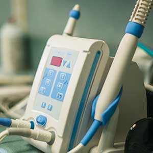
Nickel Titanium Rotary Instruments & Electric Handpieces
Nickel titanium has the unique features of flexibility and shape memory. The instruments are used to clean and shape the inside of the canal. Nickel titanium is 500 times more flexible than conventional stainless steel instruments, so even canals that are sharply curved can be thoroughly cleaned and shaped. Using these instruments routinely, allow us not only to be more effective, but also to work faster and more quietly. The electric handpieces which rotate our files make very little noise. This significantly reduces the anxiety of our patients. These handpieces are designed to be used exclusively with nickel titanium rotary instruments.
Dental Cone Beam CT Scanner
Warm Gutta Percha injection system
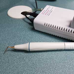
Ultrasonic Instruments
Surgical Microscope
